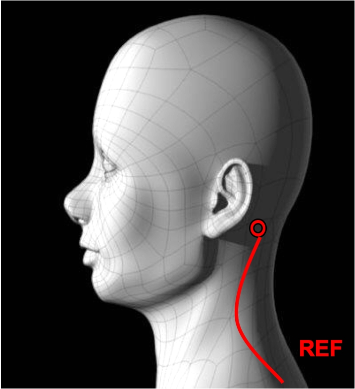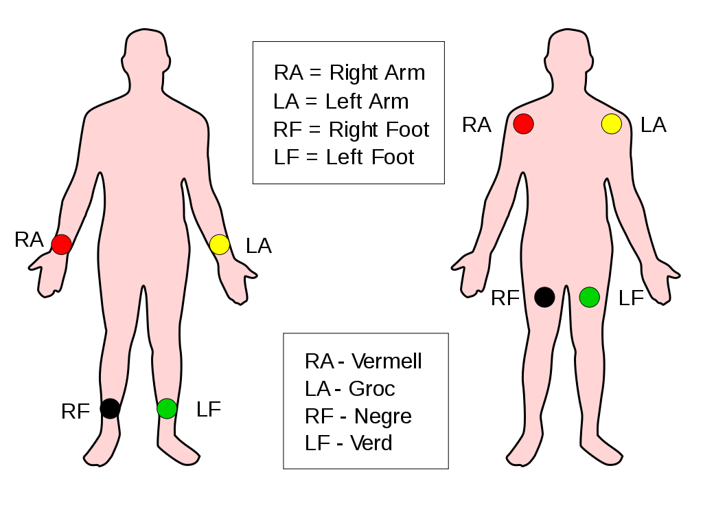


The LV summit is the most superior portion of the epicardial LV outflow tract area bounded by the left anterior descending (LAD) and left circumflex (LCx) arteries and the great cardiac vein (GCV). ... As previously described, placement of 12-lead ECG and CARTO ... Balkhy H, Tung R. Totally endoscopic robotic epicardial ablation of refractory ...
Cardiothoracic | Beebe Healthcare
•Robotic LIMA harvesting & minimally invasive Single vessel CABG •Robotic Epicardial lead placement for biventrucular pacemaker •Robotic Left Atrial Appendage Clipping or Ligation. Pericardial: •Robotic resection of Pericardial tumors (being & malignant) •Robotic resection of simple pericardial cystsIntroduction: Cardiac resynchronization therapy (CRT) is used to treat heart failure (HF) with prolonged QRS, with the LV lead typically placed transvenously (TV). Because CRT benefit correlates with concordant placement of the LV lead at the site of latest contraction, we hypothesized that epicardial (EPI) lead placement using a (robotic) surgical approach would afford more consistent access ...
Surgical approaches for epicardial LV lead placement include (1) left lateral minithoracotomy [20, 21] (2) video-assisted thoracoscopy [22], and (3) robotically-enhanced approach [21, 23, 24]. Left lateral minithoracotomy.
CONCLUSIONS: Robotic LV epicardial lead implantation results in excellent short-term response rates that persist over a 2-year follow-up and are associated with significant LV remodeling. Very robotic epicardial lv lead placement poor LV systolic function and early non-response to robotic CRT portends a poor prognosis.
Gross anatomic studies have shown a median of six veins from the left ventricle draining into the main CS. 10 The nearest branch to the CS ostium is the middle cardiac vein (MCV), which may be covered by a small valve or originate with a separate ostium. The MCV runs in the interventricular groove toward the ventricular apex and is usually not a suitable target for LV lead placement.
Using the da Vinci Robotic Surgical System (Intuitive Surgical Inc, Sunnyvale, California), a total of 19 epicardial leads were implanted successfully (1 patient received only 1 lead).
Multi-modality image fusion of 3D coronary venous anatomy from fluoroscopic venograms with left ventricular (LV) epicardial surface from single-photon emission computed tomography (SPECT) myocardial perfusion image (MPI) can provide robotic epicardial lv lead placement both LV physiological information and venous anatomy for guiding CRT LV lead placement. However, it is difficult to match the time points between MPI and …
A rare case of epicardial left ventricular sutureless ...
of an epicardial LV lead.6 Surgical epicardial LV lead placement is an established effective means of biventricular pacing when compared to coronary sinus leads.7 Figure 1 Posteroanterior and lateral chest radiograph demonstrating the position of the 2 left ventricular epicardial leads.RECENT POSTS:
- belt louis vuitton ebay
- ioffer louis vuitton cheap handbags
- homes for sale memphis tn 38115
- louis vuitton purse cleaning services
- carry on rolling luggage garment bag
- small canvas drawstring bag
- louis vuitton masters backpack
- outlet louis vuitton m41561 palm springs backpack mm monogram canvas
- garment bag luggage toughest
- dressberry backpacks online india
- lv manhattan bag reviewed
- womens leather purses on amazon
- how to verify louis vuitton date code
- louis vuitton monogram giant neverfull mm rouge

Share your thoughts