

-LV *conical in shape *smoother walls *lack of trabeculations *thicker walls *2 prominent papillary muscles *pumps O2 to body *higher pressure 120/10-RV *crescent shaped *walls are thinner *apical has trabeculations *moderator band *pumps deoxygenated blood to lungs *lower pressures 25/5
LV Non-Compaction •Left ventricular noncompaction (LVNC) is a cardiomyopathy characterized by prominent left ventricular trabeculae and deep intertrabecular prominent lv apical trabeculations on echo recesses •To be distinguished from (How?) LV hyper-trabeculation –often seen in normal •MRI required as echo insufficiently sensitive or specific…but remember, MRI ≠ TRUTH
Republished: Non-compaction cardiomyopathy | Postgraduate ...
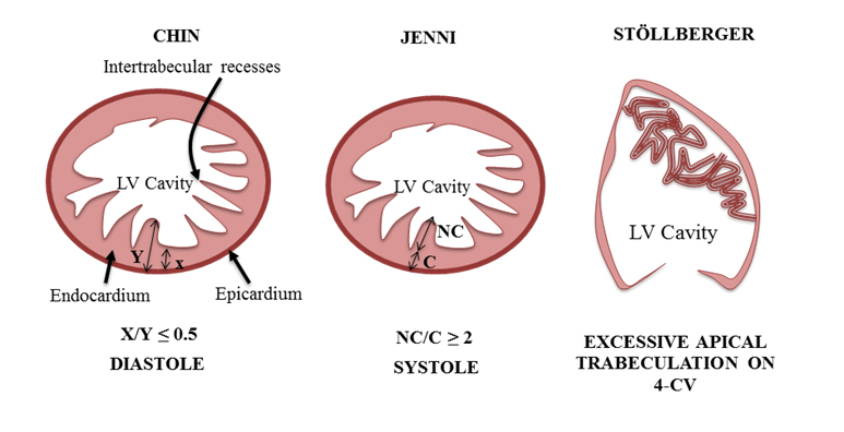
Acquisition: evaluates the trabeculations at the left ventricle (LV) apex, using the short axis and apical views and the free wall, at end-diastole. Jenni et al 3. Bilayered LV myocardium, with a thin compacted layer and a thick non-compacted layer, with trabeculations and deep recesses; ratio non-compacted/compacted >2.0The echocardiogram showed prominent trabeculations and deep intertrabecular recesses of the LV walls, especially in the posterior and apical areas. LV contrast echocardiography revealed markedly protruberant trabeculations, which were also observed with computed tomography.
Left ventricular non-compaction: A review of literature on ...
18 of gestation [23,25]. These changes are more pronounced in the LV than in the right ventricle (RV) [23,24]. For undetermined reasons, in LVNC patients, the compaction process does not occur, resulting into the development of thickened non-compacted endocardial layer having prominent diffuse trabeculations in the LV cavity. These trabeculationsMay 21, 2019 · Knowing the plane of imaging prescribed (e.g., sinotubular junction or mid-ascending aorta) is essential, as errors can occur, especially if the prominent lv apical trabeculations on echo plane is close to the annulus. Translational valve motion can produce undersampling of flow, and arrhythmias and prominent LV trabeculations …
A rare complication of acute myocardial infarction: Intra ...
An irregular ventricular wall with flow within identifies prominent trabeculations. Data on appropriate management are limited. However, studies have shown that survival at 30 days was just 10% in those managed conservatively. 6 Intra‐ventricular repair with pericardial patch, as well as Teflon patch and glue, has been used with some success ...Table 1 | The Current Approach to Diagnosis and Management ...
Description (i) Prominent trabeculations with deep recesses (ii) Decrease in ratio from MV level to papillary muscle level of the distance from the epicardium to the trough of the trabeculations to the epicardium to the prominent lv apical trabeculations on echo peak of the trabeculations ()(iii) Increasing LV wall thickness from base to apexCardiovascular Ultrasound BioMed Central
ventricle (LV) has up to 3 prominent trabeculations and is, thus, less trabeculated than the right ventricle [1,2]. Rarely, more than 3 prominent trabeculations that is the so-called LV noncompaction of ventricular myocardium (NVM) can be found at autopsy and by various imaging techniques including echocardiography and MRI etc. in the LV.RECENT POSTS:
- black supreme fanny pack fake
- louis ck girlfriend current
- louis vuitton iena pm azur
- lv twist bracelet reviewed
- how to check an authentic louis vuitton bag
- louis vuitton strap repair near memphis
- alligator skin backpack purse
- louis multi pochette accessoires
- louis vuitton monogram shine shawl grey
- meningitis belt senegal mapping
- louis vuitton perfume mens price in indiana
- replica supreme x louis vuitton hoodie
- rosalie coin purse damier azur
- levi's outlet store

Share your thoughts