

Shoulder muscles : Anatomy and functions | Kenhub
Oct 29, 2020 · Muscles of the shoulder : Anterior view. The muscles of the shoulder support and produce the movements of the shoulder girdle.They attach the appendicular skeleton of the upper limb to the axial skeleton of the trunk. Four of them are found on the anterior aspect outlet view of shoulder joint labeled of the shoulder, whereas the rest are located on the shoulder’s posterior aspect and in the back. wild bird rescue st louisCategory: Labeled-Anatomy Atlas 4E Brazil ID: 4375 Title: Shoulder (Glenohumeral) Category: Labeled-Anatomy Atlas 4E ID: 16773 Title: Hansen Flash Cards 6-4 Category: Labeled - Flash Cards
The supraspinatus outlet view reveals acromial morphologic traits, acromial slope, subacromial excrescences, and inferior osteophytes at the acromioclavicular joint. The major disadvantage of the outlet view is reproducibility. The outlet view of shoulder joint labeled purpose of this article is to describe technical factors that allow for obtaining this view consistently.
Please Note: You may not embed one of our images on your web page without a link back to our site. If you would like a large, unwatermarked image for your web page or …
Shoulder Joint Ligaments - Medical Art Library
Aug 07, 2017 · License Image The joint cavity is surrounded by a loose fitting fibrous articular capsule. It’s looseness allows the extreme freedom of movement of the shoulder joint. The capsule is strengthened by the tendons and ligaments surrounding and blending with it. The coracohumeral, glenohumeral ligaments and the tendons of the supraspinatus and subscapularis muscles all serve […]Shoulder US: Anatomy, Technique, and Scanning Pitfalls ...
Anatomy. The shoulder is a synovial articulation between the glenoid and the humeral head in which the shallow glenoid articulation is deepened an additional 50% by the fibrocartilaginous labrum that forms a rim around the perimeter of the glenoid ().Both the glenoid and the humeral head are covered by a layer of hyaline articular cartilage.SCAPULAR Y LATERAL - ANTERIOR OBLIQUE POSITION: SHOULDER ...
Apr 21, 2012 · Center scapulohumeral joint to CR and to center of IR. Abduct arm slightly if possible so as to not superimposed proximal humerus over ribs; do not attempt to rotate arm. Central Ray: CR perpendicular to IR, directed to scapulohumeral joint (2 or 2 1/2 inches [ 5 to 6 cm] below top of shoulder) see note. Minimum SID of 40 inches (100 cm ...Shoulder - Wikipedia
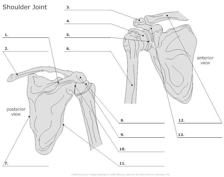
The shoulder joint (also known as the glenohumeral joint) is the main joint of the shoulder. It is a ball and socket joint that allows the arm to rotate in a circular fashion or to hinge out and up away from the body. It is formed by the articulation between the head of the humerus and the lateral scapula (specifically-the glenoid cavity of the scapula).Figure 2 (A) Grade I sprain of AC outlet view of shoulder joint labeled joint. AP view of shoulder in patient with pain and point tenderness of AC joint after fall onto shoulder demonstrates normal appearing AC joint (arrow) with no separation or fracture. (B) Grade II sprain of AC joint. AP view of shoulder demonstrates slight …
RECENT POSTS:
- lv on the go gm
- gateway arch tour st louis movie
- louis vuitton fashion show 2020
- louis vuitton bloomingdale's 59th street
- louis vuitton deutschland store
- cheap canvas sneakers
- sarah harris louis vuitton bag
- louis vuitton crossbody bag pink
- lv restaurant group
- lvmh net sales 2018
- gucci red mini backpack
- louis vuitton satchel bag price
- neverfull copy
- louis vuitton neverfull mm price usd

Share your thoughts