

Investigation of Increased LV Wall Thickness ... - echo.guru
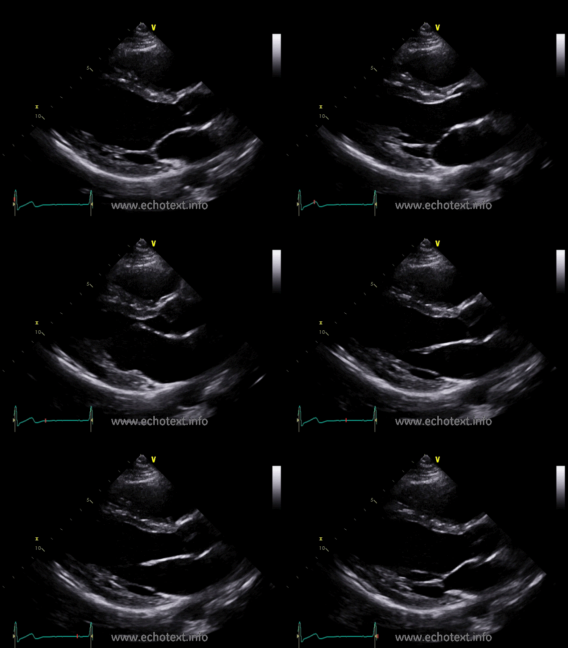
In the 3rd case you can see a normal LV cavity with only a mild degree of LV wall thickening. In fact the whole echo was fairly unremarkable other than the mild LV hypertrophy. On strain imaging you can see that overall it is normal except for a very mild reduction in regional strain at the basal septal region.Normal values for cardiovascular magnetic resonance in ...
Apr 18, 2015 · Morphological and functional parameters such as chamber size and function, aortic diameters and distensibility, flow and T1 and T2* relaxation time can be assessed and quantified by cardiovascular magnetic resonance (CMR). Knowledge of normal normal lv size by echo values for quantitative CMR is crucial to interpretation of results and to distinguish normal from disease.Small Left Ventricular Size Is an Independent Risk Factor ...
Using computational modeling and virtual surgery, we created two geometrically similar LV size models—a small LV with an LVEDd of approximately 4.5 cm and an average-sized model with an LVEDd of approximately 6.5 cm. Left ventricular assist device support in patients with small intraventricular volumes (irrespective of etiology) results in ...Size Matters! Impact of Age, Sex, Height, and Weight on ...
values are applied. The aim of the present study was to assess the impact of body size, sex, and age on the normal heart size. Methods and Results—We prospectively studied 622 individuals (52.7% women; 17–91 years; 143–200 cm; 32–240 kg) without cardiac disease by standard transthoracic echocardiography.Age and gender specific normal values of left ventricular ...
Jan 21, 2009 · Gradient echo CMR was performed at 1.5 T in 96 healthy volunteers (11–81 years, 50 male). Gender-specific analysis of parameters was undertaken in both absolute values and adjusted for body surface area (BSA). Age and gender specific normal ranges for LV volumes, mass and function are presented from the second through the eighth decade of life.Bedside Echo in Pulmonary Embolism • LITFL • Clinical ...
Nov 03, 2020 · This case illustrates the utility of bedside echocardiography in the emergency department. Using the clinical history, a diagnosis of massive pulmonary embolism was made at the bedside and appropriate treatment could be administered almost immediately. The pictures are from a real case, with some of the details changed.5 The Ventricles Pre-reading for the FCUS Course ...
A = LV internal diameter in diastole (LVIDd) = 3.7cm. B = LV internal diameter in systole (LVIDs) = 3.4cm. In this view, the LV is contracting by only 10% = gross LV hypokinesis. IS RV CONTRACTION INCREASED / DECREASED / GROSSLY NORMAL? KEY POINTS: Measurement of RV function is beyond the normal lv size by echo scope of basic cardiac echo. This is for the following ...LV Volume = [7/(2.4 + LVID)] * LVID 3. RWMA, either close normal lv size by echo or distant, may cause the volume analysis to be incorrect. If the endocardial boarder is poorly seen, then the area of …
2). LV volumes were also significantly different among the software, with some exceptions, as detailed in Table 3. The response of LVEF to vasodilator stress, a potential marker of disease severity, was relatively similar among the software (Fig. 2). Comparison of Subgroups of Healthy Subjects The mean values of normal LVEF in patients with a low gucci outlet locations
RECENT POSTS:
- louis vuitton speedy 35 diaper bag
- st louis cardinals at chicago cubs 2020
- men's leather bifold wallets made in usa
- sell my used designer handbags
- neverfull gm mm vs gmx
- lv speedy 25 damier ebene
- lv europe price 2019
- louis vuitton boots for sale scottsdale
- supreme lv dog hoodie amazon.com
- st louis blues playoff ticket prices
- bulk duffle bags cheap for football teams
- shoes louis vuitton buy online
- lv x virgil bags
- best backpacks for women

Share your thoughts