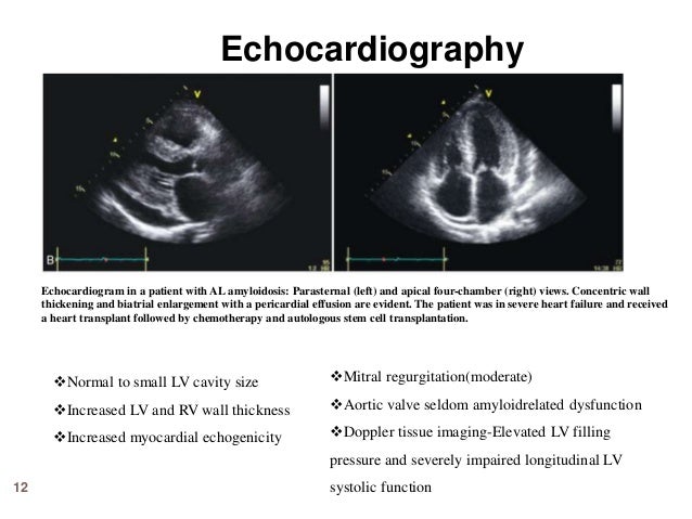


2D, Doppler, and 3D transesophageal echocardiogram performed intraoperatively for the guidance of transcatheter mitral valve repair with MitraClip®. PRE-PROCEDURE TEE: – Normal left ventricular size and systolic function. Estimated ejection fraction is 60%. – Right ventricle is mildly dilated with mildly reduced systolic function
Echocardiography - Crashing Patient
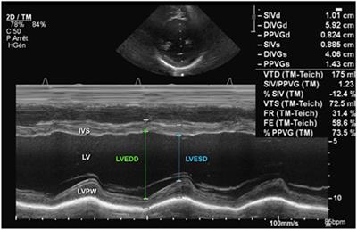
LV systolic function can be assessed using echocardiography by measuring ejection fraction (EF−normal range 55 75%), fractional shortening (FS−normal range 30 42%) and cardiac output (CO). The relevant LV dimensions can be obtained from the parasternal long axis view and EF and FS can be calculated using the formula in Box 1 .Normal values of M mode echocardiographic measurements of ...
OBJECTIVE To obtain normal M mode (one dimensional) echocardiographic values in a substantial sample of normal infants and children. DESIGN Data were obtained over three years from a single centre in central Europe. PATIENTS 2036 healthy infants and children aged one day to 18 years. METHODS In line with recommendations for standardising measurements from M mode echocardiograms, and …Echo Report Summary and Conclusions Templates | | Cordoc
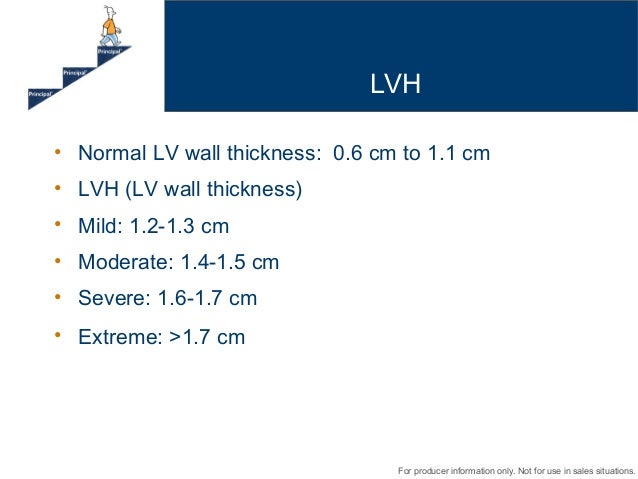
Mild concentric left ventricular hypertrophy with normal cavity size and preserved systolic function (EF 60 %) Normal right ventricular size and systolic function Sclerodegenerative valve disease with normal function. Dilated cardiomyopathy: Mildly dilated left ventricle with *** reduced systolic function.echo: normal left ventricular size + wall thickness and wall motion, normal biventricular systolic function, mild mvp w/ trace regurgitation, ef 67%, rv systolic pressure 16mmhg, mild heart murmur. no symptoms. help! is this normal?
Measurements of left ventricular dimensions using real-time acquisition in cardiac normal lv dimensions echo magnetic resonance imaging: comparison with conventional gradient echo imaging Magma: Magnetic Resonance Materials in Physics, Biology, and Medicine, Vol. 13, No. 2
As technology advances and we are encouraged to implement additional measurements into our routine scanning exams, it’s good to take a step back and refresh on proper techniques! Our goal for this blog is to have a better understanding of proper technique for measuring left ventricle (LV) volumes! In the ASE 2015 Chamber Quantification Guidelines, the […]
Impaired left ventricular relaxation
Mar 27, 2019 · Please let Kimberly know her echo looked good. Her left normal lv dimensions echo ventricle size was normal, normal wall thickness and normal ejection fraction. Wall motion was normal. It did detect possible impaired left ventricular relaxation which just means her left ventricle of her heart doesn't relax fully after each beat. This is common.Transthoracic Echocardiography in Models of Cardiac ...
LV Dimensions and Function in the Normal Mouse Heart. Representative 2D and M-mode echocardiograms in a normal mouse are shown in Fig 1. Normal mice weight-matched to AVF mice (control group 1) and those weight-matched to TGβ (control group 2) differed significantly from each other in BW because of the age difference, and there was a ...RECENT POSTS:
- zippy wallet louis vuitton reviews
- slots lv no deposit codes 2019
- louis ruelas
- supreme lv colab items
- lv m61276 ptt
- used louis vuitton bags near memphis tn
- jaden smith model louis vuitton
- amazon nylon cross body bags
- louisiana souvenirs wholesale
- louis vuitton new york store
- are louis vuitton scarves made in france
- louis vuitton laptop bags south african
- tory burch diaper bags
- outlet mall chesterfield mo black friday

Share your thoughts