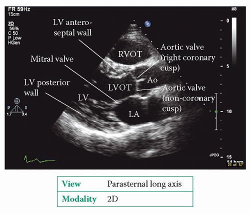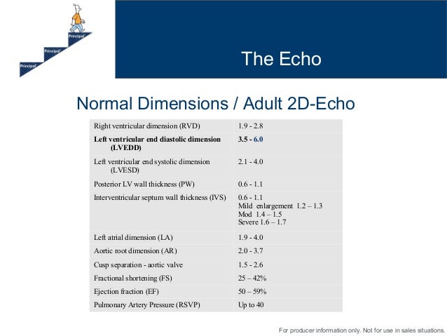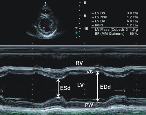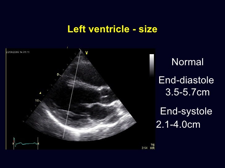


According to the American Society of Echocardiography and the European Association of Cardiovascular Imaging, diastolic dysfunction assessment on echocardiography is divided into two different groups based on left ventricular systolic function. Normal left ventricular systolic function. There are four criteria should be evaluated: average E/e ...
Measurement of left ventricular volume after anterior ...
We have compared echocardiography (echo) and radionuclide ventriculography (RNV) with magnetic resonance imaging (MRI) for the measurement of left ventricular (LV) volume and ejection fraction. Seventy asymptomatic patients were studied up to 12 days after first Q wave anterior myocardial infarction and again after 6 months.Strain measurement Left ventricle (LV) Longitudinal lv measurements echo strain is measured at the endocardial border as indicated by the green line. Instantaneous endocardial strain is visualized by color-coding close to the endocardial border. The segmental strain values are displayed on an 18-segment bull’s-eye plot. The user can select either end-systolic
OBJECTIVE To obtain normal M mode (one dimensional) echocardiographic values in a substantial sample of normal infants and children. DESIGN Data were obtained over three years from a single centre in central Europe. PATIENTS 2036 healthy infants and children aged one day to 18 years. METHODS In line with recommendations for standardising measurements from M mode echocardiograms, and …
Echocardiography tutorials
Virtual Echocardiography (Overview) LV Linear Measurements . Right Atrium Measurements . LV Mass lv measurements echo (Area-Length) LV Mass (Truncated Ellipsoid) LV Ejection Fraction, Stroke Volume, Minute Volume and Cardiac Index . Assessment of Aortic regurgitation (PHT)Comparison Between 3D Echocardiography and Cardiac ...
Background: Echocardiography and Cardiac Magnetic Resonance Imaging (CMRI) are two noninvasive techniques for the evaluation of cardiac function for patients with coronary artery diseases. Although echocardiography is the commonly used technique in clinical practice for the assessment of cardiac function, the measurement of LV volumes and left ventricular ejection fraction (LVEF) by the use of ...Sep 19, 2018 · Left ventricular lv measurements echo outflow tract gradient (LVOT) in hypertrophic obstructive cardiomyopathy (HOCM) is usually measured from the apical five chamber view (apical 5C) in echocardiography. Initially the apical 5C view is obtained and then the colour Doppler flow mapping (CFM) is done to …
Echocardiography In Heart Failure - USC Journal
M-mode echocardiographic measurements of LV function benefit from high temporal resolution, but are inaccurate in patients with segmental dysfunction or non-elliptical ventricles. Qualitative, 'eyeballÔÇÖ grading of left ventricular systolic dysfunction into mild, moderate or severe categories is widely used in clinical practice, but ...Strain rate imaging - Wikipedia
Strain rate imaging is a method in echocardiography (medical ultrasound) for measuring regional or global deformation of the myocardium (heart muscle). The term "deformation" refers to the myocardium changing shape and dimensions during the cardiac cycle. If there is myocardial ischemia, or there has been a myocardial infarction, in part of the heart muscle, this part is weakened and shows ...RECENT POSTS:
- louis vuitton looping bag strap
- neverfull gm or mm
- louis vuitton jobs in houston
- louis vuitton new wave heart bag price
- mickey mouse handbags at amazon com
- super pho menu salem
- louis vuitton lockme bucket nano bag
- cheap louis vuitton bags nz
- louis vuitton authentication service near mesa
- lv saleya damier azur
- gucci and louis vuitton airpod case
- louis vuitton branding strategy
- men's wearhouse winston salem
- best tote handbags 2018

Share your thoughts