

Hypertrophic Cardiomyopathy | Thoracic Key
Feb 26, 2019 · Other patterns also occur, including concentric, midventricular (sometimes associated with a diverticulum at the LV apex), and apical. Coexistent right ventricular hypertrophy is common but rarely, if ever, occurs in isolation. There are often associated abnormalities of the mitral valve (MV) and papillary muscles, which are also frequently ...Overview of cardiac and paracardiac aneurysms ...
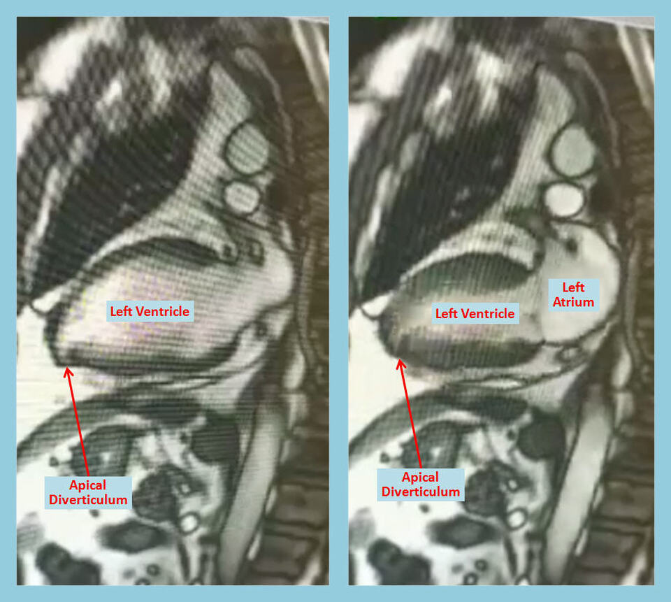
Teaching point: Ventricular diverticula are commonly found in the apex and perivalvular area, however, any segment can be involved. Apical diverticula have a high association (> 70%) with other congenital abnormalities, including septal defects, pulmonary stenosis, and dextrocardia.Subvalvular Apical Left Ventricular Aneurysms in Bantu Source
valvular and apical aneurysms are related to this typeof diverticulum. The fibrous type of congenital diverticulum of the left ventricle occurs either in the apical or the subvalvular position. Drennan lv apical diverticulum and Van der VijverI8 reported the case of a 4-year-old African female who died suddenlv following rupture of a small "cyst-like diver-Left ventricular diverticulum of the heart is a rare malfor- mation. The majority arise from the apex left ventricle and are usually found in children. Only a few cases adults have been reported. In the past, congenital aneurysm and diverticulum of the left ventricle were used synonymously. lv apical diverticulum Treisman and associates [ 11 classified the
A 69-year-old woman presenting with shortness of breath ...
A 69-year-old Caucasian woman, with a history of hypertension and type 2 diabetes mellitus, presented with exertional dyspnoea. Transthoracic echocardiography (TTE) showed moderate to severe aortic stenosis and moderate aortic regurgitation, with normal left ventricular (LV) systolic function and dimensions. Prior to referral for surgical aortic valve replacement, she underwent cardiac ...Body Surface Electrocardiographic Mapping
Jul 27, 2012 · Intracardiac lv apical diverticulum activation mapping during tachycardia demonstrated earliest activation in the apical LV diverticulum. During sinus rhythm, pace map at that site was remarkably similar to the tachycardia morphology in 12/12 ECG leads. Electroanatomic 3D mapping (CARTO) of the LV chamber performed during the ectopy also identified the earliest site ...Aug 31, 2010 · The diagnostic criterion for apical HCM is (a) an absolute apical wall thickness of more than 15 mm or (b) a ratio of apical to basal LV wall thicknesses of 1.3–1.5 (32,33).
phragm, and heart, resulting in a cardiac diverticulum [13, 181. Apical diverticula were treated as early as 1912 by reduction into the pericardium [19], and in 1944, an apical diverticulum was resected by closed ligation [20]. Epicardial cysts are a rare condition in which sinusoids occur in the myocardium. These sinusoids may or may
Ventricular aneurysm - Wikipedia
Ventricular aneurysms are one of the many complications that may occur after a heart attack.The word aneurysm refers to a bulge or ‘pocketing’ of the wall or lining of a vessel commonly occurring in the blood vessels at the base of the septum, or within the aorta.RECENT POSTS:
- craigslist st louis cars and trucks by dealer
- louis vuitton trunk ebay
- louis vuitton speedy 30 costco
- louis vuitton heels black and white
- louis vuitton lv logo font
- louis vuitton bag for sale near mesa
- louis vuitton idylle blossom
- walmart black friday 2019 ad 65 inch tv
- louisville slugger bats sale
- louis philippe brand login
- lv bags century 21
- check authentic louis vuitton neverfull
- custom supreme x louis vuitton air force 12
- louisville basketball coach list

Share your thoughts