

Transient Ischemic Attack and Ischemic Stroke in Danon ...
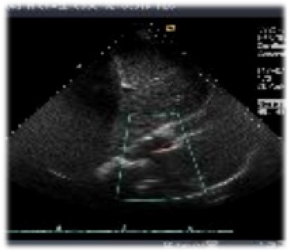
The presence of the LV apical thrombus was not detected by a conventional parasternal sweep or apical 4-chamber view but detected only by an off-axis view as we searched intensively due to the ischemic stroke (Figure 2). In the case of intracardiac thrombus in the left heart, aggressive anticoagulation should be initiated to prevent further ...Resolution of left atrial appendage thrombus with apixaban ...
Dec 20, 2013 · Left atrial appendage (LAA) thrombosis is an important cause of cardiogenic cerebral thromboembolism. Apixaban is a member of the class of novel oral anticoagulants (NOAC) and is superior to warfarin in preventing stroke or systemic embolism, causes less bleeding, and results in lower mortality in patients with atrial fibrillation. There are few reports of resolution of LAA thrombus with …Discussion. The prevalence of LV aneurysm in patients with HCM was approximately 2% in a study of 1,299 patients by Maron and colleagues. 1 However, this percentage might be an underestimate, because TTE, the imaging method most often used to diagnose HCM, can fail to detect small lv apical clot icd 10 apical aneurysms. In the same study, with the use of CMR to detect LV aneurysm, maximal wall thickness was noted ...
ICD-10-CM/PCS codes version 2016/2017/2018/2019/2020, ICD10 data search engine
Thrombosis vs. Embolism: What’s the Difference?
Nov 14, 2019 · Thrombosis and embolism share many similarities, but they are unique conditions. Thrombosis occurs when a thrombus, or blood clot, develops in a …Magnetic resonance imaging identified a calcified thrombus in the apical cavity of the LV in the setting of an atypical HCM. springer We describe a patient with a giant thrombus on the apical wall of the left ventricle that occurred due to HIT syndrome after anterior myocardial infarction.
Left ventricular apical clot resolved. Serial ECGs for 3 consecutive days displayed marked repolarization abnormalities with fluctuating prolonged QT intervals that failed to normalize. After a long discussion among all treating physicians, the electrophysiologist, and the patient, an ICD was implanted; thereafter, she was completely stable ...
Apixaban Versus Warfarin in Patients With Left Ventricular ...
Presence and dimensions of Left ventricular thrombus (LV) as assessed by 2D echocardiography [ Time Frame: lv apical clot icd 10 3 months ] The primary efficacy endpoint will be the presence of LV thrombus as assessed be echocardiography after 3 months of treatment with oral anti coagulation. Dimensions of the LV thrombus (if still present) will be compared to the ...Intra-cardiac thrombus resolution after anti-coagulation ...
Oct 08, 2013 · Thrombic progression in apical aneurysm (thrombus indicated by arrows). (A) Prior to anti-coagulation therapy, thrombus size was 15.0mm×17.0mm.(B) One week after initial dabigatran administration (150mg b.i.d.), thrombus size was 10.0mm×9.0mm.(C) Two weeks after initial dabigatran administration (150mg b.i.d.), thrombus size was 6.8mm×6.0mm.(D) Three weeks after initial …RECENT POSTS:
- black gucci coin bag
- louis l'amour free audio bookstore
- sling shoulder backpacks bags crossbody
- supreme x louis vuitton tote bags
- louis vuitton paris contact email
- vintage style wallets for women
- pm lv agenda
- louis vuitton purses speedy 35
- louis vuitton runaway sneakers
- authentic louis vuitton tags
- louis vuitton or gucci
- louis vuitton outlet store uk
- leather wallet chain for sale
- st. louis cardinals season tickets

Share your thoughts