

Curative Treatment of Electrical Storm in a Patient with ...
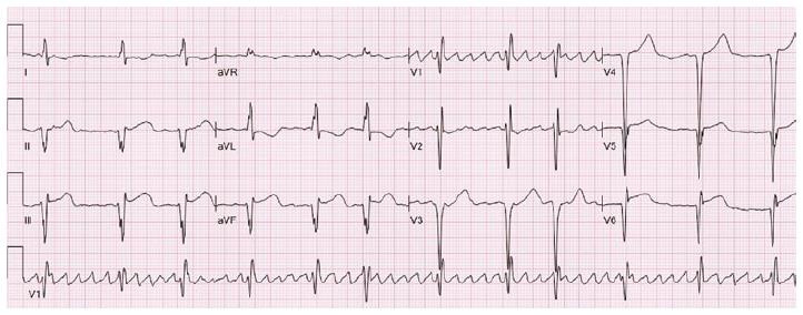
Transthoracic echocardiography disclosed moderate left ventricular systolic dysfunction, left ventricular apical aneurysm and 3 × 3 cm mural thrombus in aneurysm, which was confirmed with 3D echocardiography (Figure 2). Warfarin was added to treatment. For secondary prevention, a VVI-R ICD was implanted in our institution in January 2016.Takotsubo cardiomyopathy or Takotsubo syndrome (TTS), also known as stress cardiomyopathy, is a type of non-ischemic cardiomyopathy in which there is a sudden temporary weakening of the muscular portion of the heart. It usually appears lv apical aneurysm icd 10 after a significant stressor, either physical or emotional; when caused by the latter, the condition is sometimes called broken heart syndrome.
Mid cavity hypertrophic obstructive cardiomyopathy with ...
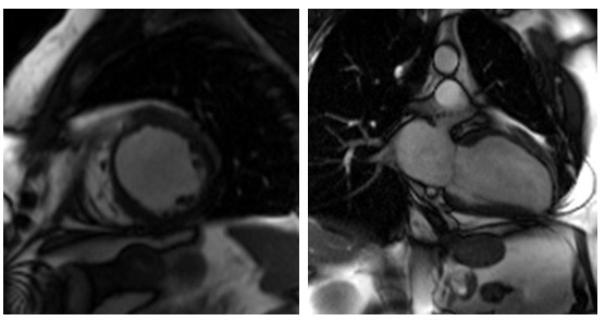
Coronary angiogram showed normal coronaries, and her left ventriculogram showed mid cavitary obliteration in systole with aneurysm of the apex (Figures 2 and 3).A cardiac MRI was performed which confirmed the diagnosis of mid-cavity hypertrophic obstructive cardiomyopathy with an apical aneurysm.INTRODUCTION: Hypertrophic Cardiomyopathy (HCM) has several distinct morphological patterns. Asymmetric left ventricular hypertrophy with outflow obstruction, mid-cavity obstruction and apical hypertrophy are types of morphological variants in HCM. Apical aneurysm formation is an uncommon feature associated with HCM. Apical aneurysms have been described in the asymmetric septal, apical, … louis vuitton sale dates
Also, LGE images revealed near full thickness scarring of the apical segments of LV (Figures 8,9,10). Figures lv apical aneurysm icd 10 5,6,7. SSFP Cines in HLA, 3 chamber, and VLA projections demonstrating apical aneurysm and mid LV thickening. Figures 8,9,10. LGE images of the LV in HLA, 3 chamber, and VLA projection showing dense scar on the mid to apical LV . Conclusion
Idiopathic left ventricular aneurysm and sudden cardiac ...
Figure 2 Left ventricular aneurysm during cardiac catheteriza-tion. The aneurysm (indicated by arrows) is depicted in the lateral LV wall in a right anterior oblique projection (RAO 308). Figure 1 Magnetic resonance imaging of an LV aneurysm. Diastolic (A) and systolic (B) imaging of the LV in …In highly selected patients with apical HCM with severe dyspnea or angina (NYHA class III or class IV) despite maximal medical therapy, and with preserved EF and small LV cavity size (LV end-diastolic volume <50 mL/m 2 and LV stroke volume <30 mL/m 2), apical myectomy by experienced surgeons at comprehensive centers may be considered to reduce ...
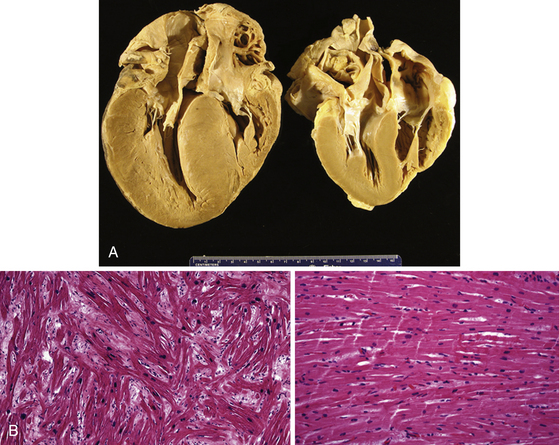
Nov 23, 2020 · He was implanted with a single-chamber implantable cardioverter defibrillator (ICD) at the age of 34. Based on the current 2010 Task Force Criteria, he has a diagnosis of definite ARVC . His last TTE showed a heavily dilated RV with reduced function and a subtricuspid aneurysm, but no LV …
CASE REPORT Open Access Epicardial unipolar …
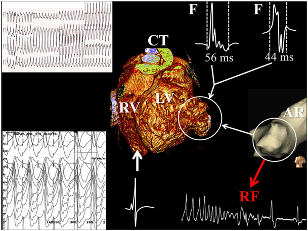
We report a case of a 62-year-old Chinese man with typical triple-vessel lesions and apical left ventricular aneurysm accompanied lv apical aneurysm icd 10 with ventricular tachycardia. Off-pump coronary artery bypass (OPCAB) grafting was performed in combination with epicardial unipolar radiofrequency ablation and linear closure of left ventricular aneurysm. TheRECENT POSTS:
- gucci marmont logo leather belt
- lv alma bb fake vs real
- louis vuitton large envelope clutch
- discount louis vuitton backpack
- mini lin louis vuitton
- speedy bandouliere 35
- louis vuitton monogram pochette clutch bag
- louis vuitton brown checkered bag
- buy diaper bag online canada
- louis vuitton leather fabric ebay
- kohl's luggage sets sale
- christmas story animation video youtube
- black friday 2019 deals appliances
- v tote mm monogram

Share your thoughts