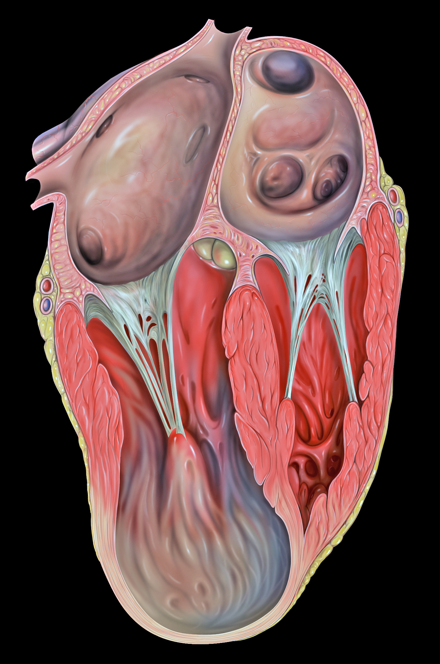


including Tako-Tsubo cardiomyopathy, LV aneurysms and pseudoaneurysms, apical diverticula, apical ventric-ular remodelling, apical hypertrophic cardiomyopathy, LV non-compaction, arrhythmogenic right ventricular dysplasia with LV involvement and LV false tendons, with an emphasis on the diagnostic criteria and imaging features.
Apical hypertrophic cardiomyopathy: what are the risks in ...
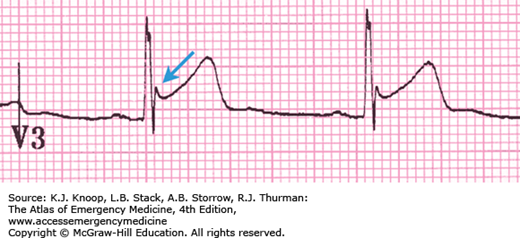
We present the case of a 50-year-old, fit, asymptomatic gurkha officer. At a routine medical, an ECG showed T-wave inversion in the chest leads V3–6. Transthoracic echo showed left ventricular apical hypertrophy and cavity obliteration consistent with apical hypertrophic cardiomyopathy (ApHCM). Cardiac magnetic resonance imaging showed apical and inferior wall hypertrophy in the left ...Cardiovascular Ultrasound BioMed Central
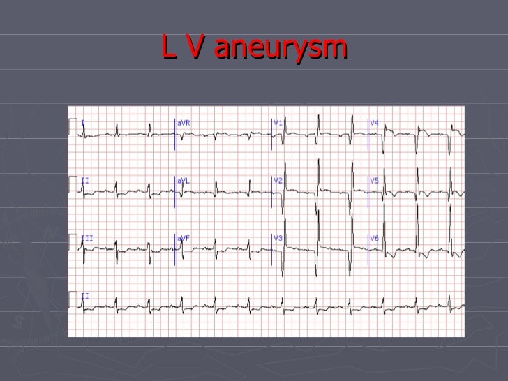
the electrocardiogram (ECG) showed a right bundle branch block (RBBB) with no Q waves or ST segment elevation. Coronary angiography revealed normal coronary arteries, left ventricular hypertrophy and an apical aneurysm. Conclusion: This case is a rare example of an …Hypertrophic cardiomyopathy | Radiology Reference Article ...
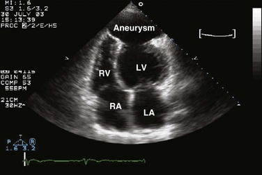
MRI is useful in evaluating HCM patients with thin-walled scarred left ventricular apical aneurysms, end-stage systolic dysfunction, massive left ventricular wall hypertrophy, associated thickening of the right ventricular wall as well as substantial morphologic diversity with regard to …A Cardiac MRI (GE Twinspeed 1.5 T) was performed lv apical aneurysm ecg to further identify the possibility of concommitant LV aneurysm and pseudoaneurysm. Steady state free precession CINE MRI demonstrated severe left ventricular dysfunction, left ventricular apical remodeling with a 22 mm collar, and thrombotic stratification (Movie 2).
Aug 03, 2020 · Left ventricular pseudoaneurysm is a potentially life-threatening complication of acute myocardial infarction. Timely diagnosis is crucial to improve the patient’s prognosis. We describe a multimodality diagnostic approach with emphasis on cardiac magnetic resonance imaging for a left ventricular pseudoaneurysm found surreptitiously in 72-year-old lv apical aneurysm ecg man 2 weeks following an acute …
Clinically suspected myocarditis with pseudoinfarct ...
apical aneurysm, with a thin, scarred myocardium (figure4). Repeated TTE confirmed the aneurysm (2.2cm width, 3.7cm length). Six months later, ECG still exhibited Q waves in anterior and inferior leads, and TTE revealed normal LV size with improvement of contrac-tility, EF 55%, but persistence of an apical aneu-rysm without thrombus.Acute Myocarditis and Left Ventricular Aneurysm as ...
In particular, atrioventricular valve incompetence and the apical LV aneurysm disappeared after 2 months of steroid therapy, while LV ejection fraction increased from 45 to 65%. Disappearance of LV aneurysm was totally unexpected, as they are considered irreversible events even when occurring as a consequence of myocardial inflammation.Intraventricular muscle bundle as a novel cause of left ...
left ventricle, which also divided the left ventricle into two distinct chambers. Blood entered the apical aneurysm in diastole and was ejected out of lv apical aneurysm ecg it in systole, as it occurs in the normal LV. The diameter of the com-munication measured 25mm in diastole. The flow rate from the main chamber to the apical aneurysm was 150 cm/s.RECENT POSTS:
- where can i buy purse supplies
- louis vuitton for unicef necklace
- bloomingdale's clearance shoes
- st louis arch cleaning video
- louis vuitton white shoe laces
- crossbody bag with chain strap cheap
- louis garneau solano 2 cycling tights (for men)
- white checkered lv purse
- lv bag collection 2018
- st louis news stations
- louis vuitton deutschland gmbh
- fendi monogram canvas bag
- plastic grocery bags with logo
- louis vuitton reverse monogram petite boite chapeau

Share your thoughts