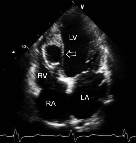


Feb 13, 2018 · We present a rare case of left ventricular (LV) lipoma. The mass measured 25 mm 10 mm, with a pedicle on the LV posterior wall near the apex. Diagnoses: The patient was diagnosed as left ventricular lipoma using echocardiography. Interventions: The LV lipoma was resected using thoracoscopy-assisted limited sternotomy. Outcomes: louis vuitton outlet
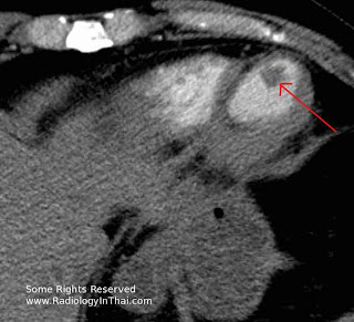
The SCT fortuitously discovered the presence of an intracardiac 2.5 cm mass, clearly delineated by contrast media, located at the level of the apex of the left ventricle. The wall thickness of the apical myocardium was also thin, suggesting an ancient left ventricular apical mass apical infarct, complicated by a thrombus.
Jun 17, 2005 · Left ventricular hypertrophy is an important risk factor in cardiovascular disease and echocardiography has been widely used for diagnosis. Although an adequate methodologic standardization exists currently, differences in measurement and interpreting data is present in most of the older clinical studies. Variability in border limits criteria, left ventricular mass formulas, body left ventricular apical mass size …
Left ventricular thrombus is a blood clot in the left ventricle of the heart. LVT is a common complication of acute myocardial infarction (AMI). Typically the clot is a mural thrombus, meaning it is on the wall of the ventricle. The primary risk of LVT is the occurrence of cardiac embolism, in which the thrombus detaches from the ventricular wall and travels through the circulation and blocks ...
Importance of length and external diameter in left ...
Background We aimed to study left ventricular (LV) geometry assessed by length (LVWL), external diameter (LVEDD) and relative wall thickness (RWT) in relation to age, body size and gender in healthy individuals. Methods 1266 individuals underwent echocardiography in the Nord-Trøndelag Health Study (HUNT3), Norway. Septum thickness (IVS), posterior wall thickness (LVPWd) and end-diastolic ...Imaging assessment of ventricular mechanics | Heart
Rotation=Rotation of short axis sections of the left ventricle as viewed from the apical end and defined as the angle (degrees or radians) between radial lines connecting left ventricular apical mass the centre of mass of that specific cross-sectional plane to a specific point in the myocardial wall …Transthoracic echocardiogram revealed normal left ventricular systolic function, with an echo-dense mass in the apex, no valvular abnormality, and mild left atrial dilatation. The patient proceeded to cardiac magnetic resonance (CMR) imaging which showed increased wall thickness in the mid to apical wall segments and high signal intensity in ...
Left ventricular regional wall motion abnormalities were noted, with apical akinesis and hypokinesis of the mid ante-rior and septal walls, as shown in the apical images displayed in Videos 1–4. A zoomed image of the apical 4-chamber view is shown in Figure 1. On the basis of the echocardio-graphic findings and patient symptoms, she was further
Differences of left ventricular systolic deformation in ...
Nov 14, 2013 · Apical hypertrophic cardiomyopathy (ApHCM), in which the myocardial wall thickening is localized at the apex of the left ventricle, is comparatively rare in western countries in comparison with Asia [1, 2].ApHCM is described as an electrocardiographic pattern of giant negative T waves and an angiographic feature of end-diastolic left ventricular cavity structure resembling an ‘ace of spades ...RECENT POSTS:
- plastic supermarket bags recyclable
- louis vuitton zippy wallet date code
- louis vuitton saintonge empreinte reviewed
- garment bags for travel australia
- best site to buy used louis vuitton
- levin furniture outlet warren oh
- why did the louisville slugger meta get banned
- six flags st. louis
- vintage 1980s louis vuitton monogram pullman suitcase
- gold chains for men cheap
- where to buy faux louis vuitton bags
- louis 90 day fiance ex wife
- black and white patent leather handbag
- outlet louis vuitton etui voyage gm m43443 monogram canvas

Share your thoughts